Explore
 Validate
 Learn
Learn Western blot
 ELISA
ELISAAntibody data
- Antibody Data
- Antigen structure
- References [0]
- Comments [0]
- Validations
- Western blot [3]
- Flow cytometry [2]
Submit
Validation data
Reference
Comment
Report error
- Product number
- NBP2-34602-0.1 mg - Provider product page

- Provider
- Novus Biologicals
- Product name
- Mouse Monoclonal TfR (Transferrin R) Antibody
- Antibody type
- Monoclonal
- Description
- Protein A purified. It recognizes a ~90-95kDa protein which is identified as cell surface transferrin receptor (CD71), a disulfide-bonded homodimeric glycoprotein of 180-190kDa (Workshop IV). This MAb is highly specific to CD71 and shows no cross-reaction with other related proteins. Ligand for transferrin receptor is the serum iron transport protein, transferrin. This receptor is broadly distributed in carcinomas, sarcomas, leukemias, and lymphomas. CD71/Transferrin receptor has been reported to be associated with cell proliferation in both normal and neoplastic tissues and useful in predicting clinical behavior or response to therapy in a number of malignancies including breast cancer.
- Reactivity
- Human
- Host
- Mouse
- Isotype
- IgG
- Vial size
- 0.1 mg
- Concentration
- 1.0 mg/ml
- Storage
- Store at 4C short term. Aliquot and store at -20C long term. Avoid freeze-thaw cycles.
No comments: Submit comment
Supportive validation
- Submitted by
- Novus Biologicals (provider)
- Main image

- Experimental details
- Simple Western: TfR (Transferrin R) Antibody (DF1513) - Azide and BSA Free [NBP2-34602] - Simple Western lane view shows a specific band for TfR (Transferrin R) in 0.2 mg/ml of Jurkat (left) and K562 (right) lysate(s). This experiment was performed under reducing conditions using the 12-230 kDa separation system.

- Submitted by
- Novus Biologicals (provider)
- Main image

- Experimental details
- Simple Western: TfR (Transferrin R) Antibody (DF1513) - Azide and BSA Free [NBP2-34602] - Electropherogram image of the corresponding Simple Western lane. TfR (Transferrin R) antibody was used at 10 ug/ml dilution of Jurkat and K562 lysates(s) respectively.

- Submitted by
- Novus Biologicals (provider)
- Main image

- Experimental details
- Western Blot: TfR (Transferrin R) Antibody (DF1513) - Azide and BSA Free [NBP2-34602] - Western Blot Analysis of Jurkat, Raji, and THP-1 cell lysate using TfR (Transferrin R) Antibody (DF1513).

Supportive validation
- Submitted by
- Novus Biologicals (provider)
- Main image
- Experimental details
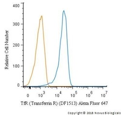
- Flow Cytometry: TfR (Transferrin R) Antibody (DF1513) - Azide and BSA Free [NBP2-34602] - A surface stain was performed on Jurkat cells with TfR (Transferrin R) Antibody (DF1513) NBP2-34602AF647 (blue) and a matched isotype control (orange).Cells were incubated in an antibody dilution of 2.5 mg/ml for 20 minutes at room temperature. Both antibodies were conjugated to Alexa Fluor 647. Image using the Alexa Fluor 647 form of this antibody.

- Submitted by
- Novus Biologicals (provider)
- Main image

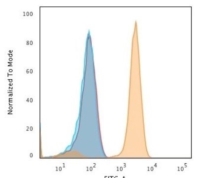
- Experimental details
- Flow Cytometry: TfR (Transferrin R) Antibody (DF1513) - Azide and BSA Free [NBP2-34602] - Flow Cytometric Analysis of human Jurkat cells using TfR (Transferrin R) Antibody (DF1513) followed by Goat anti-Mouse IgG-CF488 (Orange); cells alone (Blue); Isotype Control (Red).